World-famous cricketeer dies after booster; spike proteins destroy DNA, resulting in the coming massive “spike” in cancer

LEEDS, ENGLAND - AUGUST 25: Former Australian spin bowler Shane Warne works as a commentator for Sky Sports before the first day of the third test between England and India at Headingley on August 25, 2021 in Leeds, England. (Photo by Visionhaus/Getty Images) *** Local Caption *** Shane Warne
Spread the love

What the CNN article does not say, the Jeff Zucker-run network being infamous for its sins of omission, is that Warne attacked tennis champion Novak Djokovic for refusing to get the vaxx. Warne got the jew vaxx and the booster — and now he is dead at 52.

https://edition.cnn.com/2022/03/04/sport/shane-warne-death-spt-intl/index.html

How often have we seen celebrities bully others and virtue-signal that they got vaxxed and then keel over dead?

Instant karma.

It is the cheapest sort of bravado to speak out in total, slavish conformity with what the government, media and the Big Jews want you to say.

As Steve Kirsch reports:

The evidence

The evidence of harm has been hiding in plain sight including:

  1. An estimated reportable adverse event rate of 20% of those fully vaccinated (there are over 200M vaccinated, 1M VAERS reports and VAERS is at least 41X underreported)
  2. An estimated death due to vaccine of over 150K Americans
  3. Embalmers reporting up to 93% of cases have telltale blood clots associated with the vaccine

  4. Blood before/after vaccination is visually very different
  5. Rates of myocarditis as high as 2% (Monte Vista Christian School and a private conversation with a DoD doctor)
  6. Rates of neurological damage as high as 4.5% (Israeli MOH survey).
  7. A minimum of 30% (Peter Schirmacher’s study) to 93% (Bhakdi’s study) of deaths post vaccine attributed to the vaccine
  8. post-marketing survey disclosed by Pfizer that is pretty consistent with the VAERS data reports (I’ll write a separate substack on this later, but I didn’t see any smoking gun that isn’t already in VAERS where we already knew about thousands of adverse event types).
  9. An estimate of deaths and URF by Joel Smalley using death data in Massachusetts that confirms earlier numbers. Joel calculated a URF of 41, matching mine exactly! He also calculated a deaths per million doses (dpmd) of 945 which is even higher than the 411 dpmd calculated by Mathew Crawford.
  10. The Skidmore paper, “How Many People Died from the Covid-19 Inoculations? An Estimate Based on a Survey of the United States Population,” which estimated 294,000 excess deaths from the vaccine.
  11. German insurance company data estimate done by Mathew Crawford yielding an estimate of 120,000 deaths in the US caused by the vaccines
  12. Troubling anecdotal reports


From Natural News (https://www.naturalnews.com/2022-03-04-binary-weapon-extermination-plot-mrna-spike-protein-injections-ionizing-radiation.html?utm_referrer=https%3A%2F%2Fzen.yandex.com&utm_campaign=dbr)

The binary weapon extermination plot becomes clear: mRNA spike protein injections suppress DNA repair, followed by global nuclear events that unleash DNA-damaging radiation

This article may contain statements that reflect the opinion of the author

Bypass censorship by sharing this link:
Image: The binary weapon extermination plot becomes clear: mRNA spike protein injections suppress DNA repair, followed by global nuclear events that unleash DNA-damaging radiation

(Natural News) It is now becoming abundantly obvious what the next phase is going to be for achieving global depopulation. The vaccine bioweapon phase has achieved some level of morbid “success” in the eyes of the globalists, likely killing 1 – 2 billion human beings over the next decade as the spike protein damage takes its toll. But even this is nowhere near enough for the demonic entities controlling our planet, as they seek something closer to a 90% total reduction of the human population.

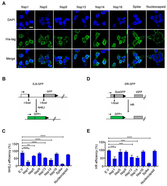
Suddenly their strategy is crystal clear. The spike protein mRNA injections cause about a 90% suppression of the DNA repair mechanism known as NHEJ – Non-Homologous End-Joining. This is a cellular mechanism that exists inside the cells of humans, animals and plants to maintain genetic integrity — a necessary condition for life.

Because of the NHEJ mechanism, we are able to automatically repair Double Strand Breaks (DSBs) in our chromosomes when we are subjected to ionizing radiation. Common sources of ionizing radiation include sunlight exposure, commercial jet flights and mammograms. When our NHEJ engine is working normally, chromosomes damaged by ionizing radiation are repaired and do not become cancer tumors. But when NHEJ is suppressed, the body cannot repair DNA damage and begins to grow micro tumors.

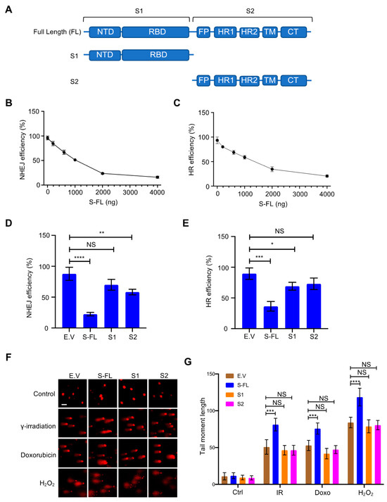
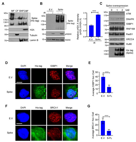
The following chart is taken from the bombshell NHEJ suppression study published in the journal VirusesYou can find it here. It shows the near-90 percent suppression of NHEJ in the presence of the spike protein which we now knows penetrates the nucleus of the cell.

Brighteon.TV 

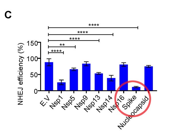
In the conclusion of the paper, the authors write, “We found that the spike protein markedly inhibited both BRCA1 and 53BP1 foci formation (Figure 3D–G). Together, these data show that the SARS–CoV–2 full–length spike protein inhibits DNA damage repair by hindering DNA repair protein recruitment.”

Micro tumors, over time — and especially in the toxic blood environment of the average vitamin D-deficient person — become large tumors. And those tumors lead to death (sometimes death by chemotherapy).

Five billion people now injected with mRNA gene therapy can be easily killed with low levels of ionizing radiation from ANY source

So right now, all across the world, there are about five billion people who have received covid vaccination injections (source) which are actually experimental gene therapy treatments that alter their DNA and suppress their DNA repair mechanisms. All that is necessary for globalists to kill those five billion people is to unleash a new source of low-level ionizing radiation that circulates across the globe… and then let physics do the rest.

Starting to get the picture of how evil these globalists really are?

This nefarious goal can be achieved by the globalists in any of the following ways:

  • Unleashing a new nuclear “accident” in Ukraine or anywhere else.
  • Setting off a nuclear bomb anywhere in the Northern hemisphere, including doing so as a false flag event to blame Russia.
  • Detonating a dirty bomb anywhere in the Northern hemisphere, potentially as an act of nuclear terrorism.

In any of these three scenarios, ionizing radiation is released and spreads across the globe due to winds. Various radioisotopes likely to be released in these events include iodine-131, cesium-137, strontium-90, plutonium-241 and others. These isotopes have various half-lives that will unleash intense iodine-131 exposure for about 10 weeks or so, followed by cesium-137 which will contaminate soils, waterways and the food supply for about 300 years (that’s about 10 half-lives). Strontium-90 has a similar half life and decay time.

No one will be able to escape the radiation exposure in the Northern hemisphere.

When mRNA vaccinated people are exposed to low levels of ionizing radiation, they will immediately start growing a swarm of new micro tumors across their entire body

A normal, healthy person with functioning NHEJ can repair ionizing radiation damage, especially if the exposure is spread out over time. But mRNA vaccinated people have lost around 90% of that repair capability. This is why mRNA-injected people are already experiencing 2000% increases in cancer rates, anecdotally reported, and it’s why we’re seeing shocking increases in all-cause mortality as reported by several life insurance providers.

The addition of a new source of global, low-level ionizing radiation would be devastating to those who have taken the mRNA injections. They would lose genetic integrity across their bodies, with tissues and organs mutating into non-functioning attempts at blood vessels and protein strands. In effect, these people’s own cells would turn against them, and it wouldn’t be long before they would suffer catastrophic failures of one or more organs or organ systems such as the circulatory system. During autopsies, it would appear as if their bodies were ravaged by a sudden wave of cancer (similar to acute radiation poisoning, but acting more slowly).

Importantly, these deaths would be diagnosed as cancer deaths, not vaccine deaths. And if a nuclear bomb could be blamed on Russia in any way, then Putin could be the scapegoat for global cancer deaths and the near-extermination of humanity.

Of course, the mRNA injections were necessary to set up the conditions for this mass die-off. Once in place, all the globalists needed was a new source of ionizing radiation to be released. And that’s fairly simple for the deep state, since Russian-made nuclear materials were smuggled out of Ukraine during the fall of the USSR in 1991, and western intelligence sources got their hands on Russian nuclear material during the chaos.

This means the globalist “deep state” has Russian nukes on hand and can set them off anywhere they want, then blame Russia for the heinous act. The obedient propaganda media of the west will gladly go along with the lie.

Similarly, Putin might actually be pushed into using his own nuclear weapons due to the extreme economic war actions that have been unleashed against Russia. We could then experience a global nuclear exchange involving several nuclear powers.

Either way, it’s clear that America and NATO are attempting to drive the world into a nuclear event of some kind, and as I’ve described in this story, I think we know the reason why. It’s the second part of the binary weapon for global depopulation.

The world is run by a suicide cult of demonic lunatics who seek the total destruction of the human race

I’ve stated this many times before, but only now are many people seeing how the dots connect. It’s now obvious that the attacks on humanity have been planned to take place in multiple vectors: Economic, biological, radiological, psychological, etc. When they are layered together on unprepared or weak-minded individuals, the result can be so devastating that their lives are quickly ended. This process is already under way and is being rapidly accelerated with the conflict in Europe. Bill Gates must be thrilled.

About 63% of the world has been injected with mRNA. Nearly 100% will be exposed to ionizing radiation as soon as the nuclear detonations (or dirty bombs) can be achieved by the globalists. 100% of the world population will face famine, starvation, fiat currency collapse and insane fuel prices due to the economic war that has recently been unleashed. And of course, nearly 100% of the population is subjected to the psychological terrorism of the lying corporate media and government propaganda.

Not surprisingly, very few individuals can withstand all of this with their minds and bodies intact. Count your blessings, because you are among these very few people.

Understand, though, that if the globalists achieve their desired escalation, many of the people you know will be dead within two years. They will die from “cancer,” from famine, suicide, lawless violence, immunological disorders, etc. Very few people who did not prepare will survive what is coming. For the most part, only the prepared will have a chance of making it through.

Article

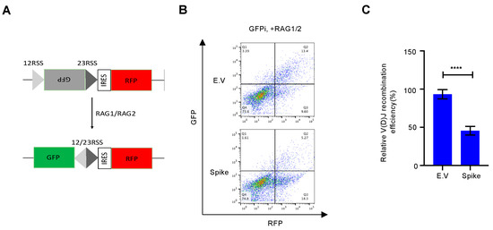
SARS–CoV–2 Spike Impairs DNA Damage Repair and Inhibits V(D)J Recombination In Vitro

by

 1,2,* and

 2,*

[source:https://www.mdpi.com/1999-4915/13/10/2056/htm]
1
Department of Molecular Biosciences, The Wenner–Gren Institute, Stockholm University, SE-10691 Stockholm, Sweden
2
Department of Clinical Microbiology, Virology, Umeå University, SE-90185 Umeå, Sweden
*
Authors to whom correspondence should be addressed.
Academic Editor: Oliver Schildgen
Viruses 202113(10), 2056; https://doi.org/10.3390/v13102056
Received: 20 August 2021 / Revised: 8 September 2021 / Accepted: 8 October 2021 / Published: 13 October 2021
(This article belongs to the Special Issue SARS-CoV-2 Host Cell Interactions)

Abstract

Severe acute respiratory syndrome coronavirus 2 (SARS–CoV–2) has led to the coronavirus disease 2019 (COVID–19) pandemic, severely affecting public health and the global economy. Adaptive immunity plays a crucial role in fighting against SARS–CoV–2 infection and directly influences the clinical outcomes of patients. Clinical studies have indicated that patients with severe COVID–19 exhibit delayed and weak adaptive immune responses; however, the mechanism by which SARS–CoV–2 impedes adaptive immunity remains unclear. Here, by using an in vitro cell line, we report that the SARS–CoV–2 spike protein significantly inhibits DNA damage repair, which is required for effective V(D)J recombination in adaptive immunity. Mechanistically, we found that the spike protein localizes in the nucleus and inhibits DNA damage repair by impeding key DNA repair protein BRCA1 and 53BP1 recruitment to the damage site. Our findings reveal a potential molecular mechanism by which the spike protein might impede adaptive immunity and underscore the potential side effects of full-length spike-based vaccines.

1. Introduction

Severe acute respiratory syndrome coronavirus 2 (SARS–CoV–2) is responsible for the ongoing coronavirus disease 2019 (COVID–19) pandemic that has resulted in more than 2.3 million deaths. SARS–CoV–2 is an enveloped single positive–sense RNA virus that consists of structural and non–structural proteins [1]. After infection, these viral proteins hijack and dysregulate the host cellular machinery to replicate, assemble, and spread progeny viruses [2]. Recent clinical studies have shown that SARS–CoV–2 infection extraordinarily affects lymphocyte number and function [3,4,5,6]. Compared with mild and moderate survivors, patients with severe COVID–19 manifest a significantly lower number of total T cells, helper T cells, and suppressor T cells [3,4]. Additionally, COVID–19 delays IgG and IgM levels after symptom onset [5,6]. Collectively, these clinical observations suggest that SARS–CoV–2 affects the adaptive immune system. However, the mechanism by which SARS–CoV–2 suppresses adaptive immunity remains unclear.
As two critical host surveillance systems, the immune and DNA repair systems are the primary systems that higher organisms rely on for defense against diverse threats and tissue homeostasis. Emerging evidence indicates that these two systems are interdependent, especially during lymphocyte development and maturation [7]. As one of the major double-strand DNA break (DSB) repair pathways, non-homologous end joining (NHEJ) repair plays a critical role in lymphocyte–specific recombination–activating gene endonuclease (RAG) –mediated V(D)J recombination, which results in a highly diverse repertoire of antibodies in B cell and T cell receptors (TCRs) in T cells [8]. For example, loss of function of key DNA repair proteins such as ATM, DNA–PKcs, 53BP1, et al., leads to defects in the NHEJ repair which inhibit the production of functional B and T cells, leading to immunodeficiency [7,9,10,11]. In contrast, viral infection usually induces DNA damage via different mechanisms, such as inducing reactive oxygen species (ROS) production and host cell replication stress [12,13,14]. If DNA damage cannot be properly repaired, it will contribute to the amplification of viral infection-induced pathology. Therefore, we aimed to investigate whether SARS–CoV–2 proteins hijack the DNA damage repair system, thereby affecting adaptive immunity in vitro.

2. Materials and Methods

2.1. Antibodies and Reagents

DAPI (Cat #MBD0015), doxorubicin (Cat #D1515), H2O2 (Cat #H1009), and β-tubulin antibodies (Cat #T4026) were purchased from Sigma-Aldrich. Antibodies against His tag (Cat #12698), H2A (Cat #12349), H2A.X (Cat #7631), γ–H2A.X (Cat #2577), Ku80 (Cat # 2753), and Rad51(Cat #8875) were purchased from Cell Signaling Technology (Danvers, MA, USA). 53BP1(Cat #NB100-304) and RNF168 (Cat #H00165918–M01) antibodies were obtained from Novus Biologicals (Novus Biologicals, Littleton, CO, USA). Lamin B (Cat #sc–374015), ATM (Cat #sc–135663), DNA–PK (Cat #sc–5282), and BRCA1(Cat #sc–28383) antibodies were purchased from Santa Cruz Biotechnology (Santa Cruz, CA, USA). XRCC4 (Cat #PA5–82264) antibody was purchased from Thermo Fisher Scientific (Waltham, MA, USA).

2.2. Plasmids

pHPRT–DRGFP and pCBASceI were kindly gifted by Maria Jasin (Addgene plasmids #26476 and #26477) [15]. pimEJ5GFP was a gift from Jeremy Stark (Addgene plasmid #44026) [16]. The NSP1, NSP9, NSP13, NSP14, NSP16, spike, and nucleocapsid proteins were first synthesized with codon optimization and then cloned into a mammalian expression vector pUC57 with a C–terminal 6xHis tag. A 12–spacer RSS–GFP inverted complementary sequence–a 23–spacer RSS was synthesized for the V(D)J reporter vector. Then, the sequence was cloned into the pBabe–IRES–mRFP vector to generate the pBabe–12RSS–GFPi–23RSS–IRES–mRFP reporter vector. 12–spacer RSS sequence: 5′–CACAGTGCTACAGACTGGAACAAAAACC–3′. 23–spacer RSS sequence: 5′–CACAGTGGTAGTACTCCACTGTCTGGCTGTACAAAAACC–3′. RAG1 and RAG2 expression constructs were generously gifted by Martin Gellert (Addgene plasmid #13328 and #13329) [17].

2.3. Cells and Cell Culture

HEK293T and HEK293 cells obtained from the American Type Culture Collection (ATCC) were cultured under 5% CO2 at 37 °C in Dulbecco’s modified Eagle’s medium (DMEM, high glucose, GlutaMAX) (Life Technologies, Carlsbad, CA, USA) containing 10% (v/v) fetal calf serum (FCS, Gibco), 1% (v/v) penicillin (100 IU/mL), and streptomycin (100 μg/mL). HEK293T–DR–GFP and HEK293T–EJ5–GFP reporter cells were generated as previously described and cultured under 5% CO2 at 37 °C in the above-mentioned culture medium.

2.4. HR and NHEJ Reporter Assays

HR and NHEJ repair in HEK293T cells were measured as described previously using DR–GFP and EJ5–GFP stable cells. Briefly, 0.5 × 10⁶ HEK293T stable reporter cells were seeded in 6–well plates and transfected with 2 μg I–SceI expression plasmid (pCBASceI) together with SARS–CoV–2 proteins expression plasmids. Forty–eight hours post–transfection and aspirin treatment, cells were harvested and analyzed by flow cytometry analysis for GFP expression. The means were obtained from three independent experiments.

2.5. Cellular Fractionation and Immunoblotting

For the cellular fraction assay, the Subcellular Protein Fractionation Kit (Thermo Fisher) was used according to the manufacturer’s instructions. Protein lysates were quantified using the BCA reagent (Thermo Fisher Scientific, Rockford, IL, USA). Proteins were resolved by sodium dodecyl sulfate–polyacrylamide gel electrophoresis (SDS–PAGE), transferred to nitrocellulose membranes (Amersham protran, 0.45 μm NC), and immunoblotted with specific primary antibodies followed by HRP–conjugated secondary antibodies. Protein bands were detected using SuperSignal West Pico or Femto Chemiluminescence kit (Thermo Fisher Scientific).

2.6. Comet Assay

Cells were treated with different DNA damage reagents and then harvested at the indicated time points for analysis. Cells (1 × 10⁵ cells/mL in cold phosphate-buffered saline [PBS]) were resuspended in 1% low–melting agarose at 40 °C at a ratio of 1:3 vol/vol and pipetted onto a CometSlide. Slides were then immersed in prechilled lysis buffer (1.2 M NaCl, 100 mM EDTA, 0.1% sodium lauryl sarcosinate, 0.26 M NaOH pH > 13) for overnight (18–20 h) lysis at 4 °C in the dark. Slides were then carefully removed and submerged in rinse buffer (0.03 M NaOH and 2 mM EDTA, pH > 12) at room temperature (RT) for 20 min in the dark. This washing step was repeated twice. The slides were transferred to a horizontal electrophoresis chamber containing rinse buffer and separated for 25 min at a voltage of 0.6 V/cm. Finally, the slides were washed with distilled water, stained with 10 μg/mL propidium iodide, and analyzed by fluorescence microscopy. Twenty fields with approximately 100 cells in each sample were evaluated and quantified using the Fiji software to determine the tail length (tail moment).

2.7. Immunofluorescence

Cells were seeded on glass coverslips in a 12–well plate and transfected with the indicated plasmid for 24 h. Then, the cells were treated with or without DNA damage reagents according to the experimental setup. The cells were fixed in 4% paraformaldehyde (PFA) in PBS for 20 min at RT and then permeabilized in 0.5% Triton X–100 for 10 min. Slides were blocked in 5% normal goat serum (NGS) and incubated with primary antibodies diluted in 1% NGS overnight at 4 °C. Samples were then incubated with the indicated secondary antibodies labeled with Alexa Fluor 488 or 555 (Invitrogen) diluted in 1% NGS at RT for 1 h. Thereafter, they were stained with DAPI for 15 min at RT. Coverslips were mounted using Dako Fluorescence Mounting Medium (Agilent) and imaged using a Nikon confocal microscope (Eclipse C1 Plus). All scoring was performed under blinded conditions.

2.8. Analysis of V(D)J Recombination

Briefly, V(D)J reporter plasmid contains inverted-GFP and IRES driving continuously expressed RFP. Continuously expressed RFP is the internal transfection control. After Recombination activation gene1/2 (RAG1/2) co–transfected into the cells, RAG1/2 will cut the RSS and mediated induction of DSBs, if V(D)J recombination occurs, the inverted GFPs are ligated in positive order by NHEJ repair. Then the cell will express functional GFP. So, the GFP and RFP double positive cells are the readout of the V(D)J reporter assay [18]. 293T cells at 70% confluency were transfected with the V(D)J GFP reporter alone (background) or in combination with RAG1 and RAG2 expression constructs, at a ratio of 1 µg V(D)J GFP reporter: 0.5 µg RAG1: 0.5 µg RAG2. The following day, the medium was changed, and after an additional 48 h, cells were harvested and analyzed by flow cytometry for GFP and RFP expression.

2.9. Statistical Analysis

All experiments were repeated at least three times using independently collected or prepared samples. Data were analyzed by Student’s t test or ANOVA followed by Tukey’s multiple-comparison tests using GraphPad 8.

3. Results

3.1. Effect of Nuclear–Localized SARS–CoV–2 Viral Proteins on DNA Damage Repair

DNA damage repair occurs mainly in the nucleus to ensure genome stability. Although SARS–CoV–2 proteins are synthesized in the cytosol [1], some viral proteins are also detectable in the nucleus, including Nsp1, Nsp5, Nsp9, Nsp13, Nsp14, and Nsp16 [19]. We investigated whether these nuclear-localized SARS–CoV–2 proteins affect the host cell DNA damage repair system. For this, we constructed these viral protein expression plasmids together with spike and nucleoprotein expression plasmids, which are generally considered cytosol–localized proteins. We confirmed their expression and localization by immunoblotting and immunofluorescence (Figure 1A and Figure S1A). Our results were consistent with those from previous studies [19]; Nsp1, Nsp5, Nsp9, Nsp13, Nsp14, and Nsp16 proteins are indeed localized in the nucleus, and nucleoproteins are mainly localized in the cytosol. Surprisingly, we found the abundance of the spike protein in the nucleus (Figure 1A). NHEJ repair and homologous recombination (HR) repair are two major DNA repair pathways that not only continuously monitor and ensure genome integrity but are also vital for adaptive immune cell functions [9]. To evaluate whether these viral proteins impede the DSB repair pathway, we examined the repair of a site-specific DSB induced by the I–SceI endonuclease using the direct repeat–green fluorescence protein (DR–GFP) and the total-NHEJ-GFP (EJ5–GFP) reporter systems for HR and NHEJ, respectively [15,16]. Overexpression of Nsp1, Nsp5, Nsp13, Nsp14, and spike proteins diminished the efficiencies of both HR and NHEJ repair (Figure 1B–E and Figure S2A,B). Moreover, we also found that Nsp1, Nsp5, Nsp13, and Nsp14 overexpression dramatically suppressed proliferation compared with other studied proteins (Figure S3A,B). Therefore, the inhibitory effect of Nsp1, Nsp5, Nsp13, and Nsp14 on DNA damage repair may be due to secondary effects, such as growth arrest and cell death. Interestingly, overexpressed spike protein did not affect cell morphology or proliferation but significantly suppressed both HR and NHEJ repair (Figure 1B–E, Figures S2A,B and S3A,B).
Figure 1. Effect of severe acute respiratory syndrome coronavirus 2 (SARS–CoV–2) nuclear-localized proteins on DNA damage repair. (A) Subcellular distribution of the SARS–CoV–2 proteins. Immunofluorescence was performed at 24 h after transfection of the plasmid expressing the viral proteins into HEK293T cells. Scale bar: 10 µm. (B) Schematic of the EJ5-GFP reporter used to monitor non-homologous end joining (NHEJ). (C) Effect of empty vector (E.V) and SARS–CoV–2 proteins on NHEJ DNA repair. The values represent the mean ± standard deviation (SD) from three independent experiments (see representative FACS plots in Figure S2A). (D) Schematic of the DR-GFP reporter used to monitor homologous recombination (HR). (E) Effect of E.V and SARS–CoV–2 proteins on HR DNA repair. The values represent the mean ± SD from three independent experiments (see representative FACS plots in Figure S2B). The values represent the mean ± SD, n = 3. Statistical significance was determined using one-way analysis of variance (ANOVA) in (C,E). ** p < 0.01, *** p < 0.001, **** p <  0.0001.

3.2. SARS–CoV–2 Spike Protein Inhibits DNA Damage Repair

Because spike proteins are critical for mediating viral entry into host cells and are the focus of most vaccine strategies [20,21], we further investigated the role of spike proteins in DNA damage repair and its associated V(D)J recombination. Spike proteins are usually thought to be synthesized on the rough endoplasmic reticulum (ER) [1]. After posttranslational modifications such as glycosylation, spike proteins traffic via the cellular membrane apparatus together with other viral proteins to form the mature virion [1]. Spike protein contains two major subunits, S1 and S2, as well as several functional domains or repeats [22] (Figure 2A). In the native state, spike proteins exist as inactive full–length proteins. During viral infection, host cell proteases such as furin protease activate the S protein by cleaving it into S1 and S2 subunits, which is necessary for viral entry into the target cell [23]. We further explored different subunits of the spike protein to elucidate the functional features required for DNA repair inhibition. Only the full–length spike protein strongly inhibited both NHEJ and HR repair (Figure 2B–E and Figure S4A,B). Next, we sought to determine whether the spike protein directly contributes to genomic instability by inhibiting DSB repair. We monitored the levels of DSBs using comet assays. Following different DNA damage treatments, such as γ–irradiation, doxorubicin treatment, and H2O2 treatment, there is less repair in the presence of the spike protein (Figure 2F,G). Together, these data demonstrate that the spike protein directly affects DNA repair in the nucleus.
Figure 2. Severe acute respiratory syndrome coronavirus 2 (SARS–CoV–2) spike protein inhibits DNA damage repair. (A) Schematic of the primary structure of the SARS–CoV–2 spike protein. The S1 subunit includes an N–terminal domain (NTD, 14–305 residues) and a receptor–binding domain (RBD, 319–541 residues). The S2 subunit consists of the fusion peptide (FP, 788–806 residues), heptapeptide repeat sequence 1 (HR1, 912–984 residues), HR2 (1163–1213 residues), TM domain (TM, 1213–1237 residues), and cytoplasm domain (CT,1237–1273 residues). (B,C) Effect of titrated expression of the spike protein on DNA repair in HEK–293T cells. (D,E) Only full-length spike protein inhibits non-homologous end joining (NHEJ) and homologous recombination (HR) DNA repair. The values represent the mean ± SD from three independent experiments (see representative FACS plots in Figure S4A,B). (F) Full–length spike (S–FL) protein–transfected HEK293T cells exhibited more DNA damage than empty vector-, S1–, and S2–transfected cells under different DNA damage conditions. For doxorubicin: 4 µg/mL, 2 h. For γ–irradiation: 10 Gy, 30 min. For H2O2: 100 µM, 1 h. Scale bar: 50 µm. (G) Corresponding quantification of the comet tail moments from 20 different fields with n > 200 comets of three independent experiments. Statistical significance was assessed using a two-way analysis of variance (ANOVA). NS (Not Significant): * p > 0.05, ** p < 0.01, *** p < 0.001, **** p < 0.0001.

3.3. Spike Proteins Impede the Recruitment of DNA Damage Repair Checkpoint Proteins

To confirm the existence of spike protein in the nucleus, we performed subcellular fraction analysis and found that spike proteins are not only enriched in the cellular membrane fraction but are also abundant in the nuclear fraction, with detectable expression even in the chromatin–bound fraction (Figure 3A). We also observed that the spike has three different forms, the higher band is a highly glycosylated spike, the middle one is a full–length spike, and the lower one is a cleaved spike subunit. Consistent with the comet assay, we also found the upregulation of the DNA damage marker, γ–H2A.X, in spike protein–overexpressed cells under DNA damage conditions (Figure 3B). A recent study suggested that spike proteins induce ER stress and ER–associated protein degradation [24]. To exclude the possibility that the spike protein inhibits DNA repair by promoting DNA repair protein degradation, we checked the expression of some essential DNA repair proteins in NHEJ and HR repair pathways and found that these DNA repair proteins were stable after spike protein overexpression (Figure 3C). To determine how the spike protein inhibits both NHEJ and HR repair pathways, we analyzed the recruitment of BRCA1 and 53BP1, which are the key checkpoint proteins for HR and NHEJ repair, respectively. We found that the spike protein markedly inhibited both BRCA1 and 53BP1 foci formation (Figure 3D–G). Together, these data show that the SARS–CoV–2 full–length spike protein inhibits DNA damage repair by hindering DNA repair protein recruitment.
Figure 3. Severe acute respiratory syndrome coronavirus 2 (SARS–CoV–2) spike protein impedes the recruitment of DNA damage repair checkpoint proteins. (A) Membrane fraction (MF), cytosolic fraction (CF), soluble nuclear fraction (SNF), and chromatin-bound fraction (CBF) from HEK293T cells transfected with SARS–CoV–2 spike protein were immunoblotted for His-tag spike and indicated proteins. (B) Left: Immunoblots of DNA damage marker γH2AX in empty vector (E.V)– and spike protein–expressing HEK293T cells after 10 Gy γ-irradiation. Right: corresponding quantification of immunoblots in left. The values represent the mean ± SD (n = 3). Statistical significance was determined using Student’s t-test. **** p < 0.0001. (C) Immunoblots of DNA damage repair related proteins in spike protein–expressing HEK293T cells. (D) Representative images of 53BP1 foci formation in E.V– and spike protein-expressing HEK293 cells exposed to 10 Gy γ–irradiation. Scale bar: 10 µm. (E) Quantitative analysis of 53BP1 foci per nucleus. The values represent the mean ± SEM, n = 50. (F) BRCA1 foci formation in empty vector- and spike protein-expressing HEK293 cells exposed to 10 Gy γ–irradiation. Scale bar: 10 µm. (G). Quantitative analysis of BRCA1 foci per nucleus. The values represent the mean ± SEM, n = 50. Statistical significance was determined using Student’s t-test. **** p < 0.0001.

3.4. Spike Protein Impairs V(D)J Recombination In vitro

DNA damage repair, especially NHEJ repair, is essential for V(D)J recombination, which lies at the core of B and T cell immunity [9]. To date, many approved SARS–CoV–2 vaccines, such as mRNA vaccines and adenovirus–COVID–19 vaccines, have been developed based on the full–length spike protein [25]. Although it is debatable whether SARS–CoV–2 directly infects lymphocyte precursors [26,27], some reports have shown that infected cells secrete exosomes that can deliver SARS–CoV–2 RNA or protein to target cells [28,29]. We further tested whether the spike protein reduced NHEJ–mediated V(D)J recombination. For this, we designed an in vitro V(D)J recombination reporter system according to a previous study [18] (Figure S5). Compared with the empty vector, spike protein overexpression inhibited RAG–mediated V(D)J recombination in this in vitro reporter system (Figure 4).
Figure 4. Spike protein impairs V(D)J recombination in vitro. (A) Schematic of the V(D)J reporter system. (B) Representative plots of flow cytometry show that the SARS–CoV–2 spike protein impedes V(D)J recombination in vitro. (C) Quantitative analysis of relative V(D)J recombination. The values represent the mean ± SD, n = 3. Statistical significance was determined using Student’s t-test. **** p < 0.0001.

4. Discussion

Our findings provide evidence of the spike protein hijacking the DNA damage repair machinery and adaptive immune machinery in vitro. We propose a potential mechanism by which spike proteins may impair adaptive immunity by inhibiting DNA damage repair. Although no evidence has been published that SARS–CoV–2 can infect thymocytes or bone marrow lymphoid cells, our in vitro V(D)J reporter assay shows that the spike protein intensely impeded V(D)J recombination. Consistent with our results, clinical observations also show that the risk of severe illness or death with COVID–19 increases with age, especially older adults who are at the highest risk [22]. This may be because SARS–CoV–2 spike proteins can weaken the DNA repair system of older people and consequently impede V(D)J recombination and adaptive immunity. In contrast, our data provide valuable details on the involvement of spike protein subunits in DNA damage repair, indicating that full–length spike–based vaccines may inhibit the recombination of V(D)J in B cells, which is also consistent with a recent study that a full–length spike–based vaccine induced lower antibody titers compared to the RBD–based vaccine [28]. This suggests that the use of antigenic epitopes of the spike as a SARS–CoV–2 vaccine might be safer and more efficacious than the full–length spike. Taken together, we identified one of the potentially important mechanisms of SARS–CoV–2 suppression of the host adaptive immune machinery. Furthermore, our findings also imply a potential side effect of the full–length spike–based vaccine. This work will improve the understanding of COVID–19 pathogenesis and provide new strategies for designing more efficient and safer vaccines.

Supplementary Materials

The following are available online at https://www.mdpi.com/article/10.3390/v13102056/s1, Figure S1: Expression of nuclear–localized SARS–CoV–2 proteins in human cells, Figure S2: Effect of nuclear SARS–CoV–2 proteins on NHEJ– and HR–DNA repair pathway, Figure S3: Nsp1, Nsp5, Nsp13, Nsp14 but not spike inhibit cell proliferation, Figure S4: Effect of SARS–CoV–2 spike mutants on NHEJ– and HR– DNA repair pathway, Figure S5: In vitro V(D)J recombination assay.

Author Contributions

H.J. conceived and designed the study. H.J. and Y.-F.M. supervised the study, performed experiments, and interpreted the data. Writing—original draft preparation, H.J.; Writing—review and editing, H.J. and Y.-F.M.; funding acquisition, Y.-F.M. All authors have read and agreed to the published version of the manuscript.

Funding

This work was supported by Umeå University, Medical Faculty’s Planning grants for COVID–19 (research project number: 3453 16032 to Y.F.M.); the Lion’s Cancer Research Foundation at Umeå University (grants: LP 17–2153, AMP 19–982, and LP 20–2256 to Y.F.M.), and the base unit’s ALF funds for research at academic healthcare units and university healthcare units in the northern healthcare region (ALF–Basenheten: 2019, 2020, 2021 to Y.F.M.).

Institutional Review Board Statement

Not applicable, because of this study not involving humans or animals.

Informed Consent Statement

Not applicable, because of this study not involving humans.

Data Availability Statement

The data presented in this study are available in the main text and Supplementary Materials.

Conflicts of Interest

The authors have declared that no competing interests exist. The funders had no role in study design, data collection and analysis, decision to publish, or preparation of the manuscript.

References

  1. V’Kovski, P.; Kratzel, A.; Steiner, S.; Stalder, H.; Thiel, V. Coronavirus biology and replication: Implications for SARS-CoV-2. Nat. Rev. Microbiol. 202119, 155–170. [Google Scholar] [CrossRef]
  2. Suryawanshi, R.K.; Koganti, R.; Agelidis, A.; Patil, C.D.; Shukla, D. Dysregulation of Cell Signaling by SARS-CoV-2. Trends Microbiol. 202129, 224–237. [Google Scholar] [CrossRef]
  3. Qin, C.; Zhou, L.; Hu, Z.; Zhang, S.; Yang, S.; Tao, Y.; Xie, C.; Ma, K.; Shang, K.; Wang, W.; et al. Dysregulation of Immune Response in Patients With Coronavirus 2019 (COVID-19) in Wuhan, China. Clin. Infect. Dis. 202071, 762–768. [Google Scholar] [CrossRef] [PubMed]
  4. Wang, F.; Nie, J.; Wang, H.; Zhao, Q.; Xiong, Y.; Deng, L.; Song, S.; Ma, Z.; Mo, P.; Zhang, Y. Characteristics of Peripheral Lymphocyte Subset Alteration in COVID-19 Pneumonia. J. Infect. Dis. 2020221, 1762–1769. [Google Scholar] [CrossRef] [PubMed]
  5. Zhang, G.; Nie, S.; Zhang, Z.; Zhang, Z. Longitudinal Change of Severe Acute Respiratory Syndrome Coronavirus 2 Antibodies in Patients with Coronavirus Disease 2019. J. Infect. Dis. 2020222, 183–188. [Google Scholar] [CrossRef] [PubMed]
  6. Long, Q.X.; Liu, B.Z.; Deng, H.J.; Wu, G.C.; Deng, K.; Chen, Y.K.; Liao, P.; Qiu, J.F.; Lin, Y.; Cai, X.F.; et al. Antibody responses to SARS-CoV-2 in patients with COVID-19. Nat. Med. 202026, 845–848. [Google Scholar] [CrossRef]
  7. Bednarski, J.J.; Sleckman, B.P. Lymphocyte development: Integration of DNA damage response signaling. Adv. Immunol. 2012116, 175–204. [Google Scholar] [PubMed]
  8. Malu, S.; Malshetty, V.; Francis, D.; Cortes, P. Role of non-homologous end joining in V(D)J recombination. Immunol. Res. 201254, 233–246. [Google Scholar] [CrossRef]
  9. Bednarski, J.J.; Sleckman, B.P. At the intersection of DNA damage and immune responses. Nat. Rev. Immunol. 201919, 231–242. [Google Scholar] [CrossRef]
  10. Gapud, E.J.; Sleckman, B.P. Unique and redundant functions of ATM and DNA-PKcs during V(D)J recombination. Cell. Cycle 201110, 1928–1935. [Google Scholar] [CrossRef]
  11. Difilippantonio, S.; Gapud, E.; Wong, N.; Huang, C.Y.; Mahowald, G.; Chen, H.T.; Kruhlak, M.J.; Callen, E.; Livak, F.; Nussenzweig, M.C.; et al. 53BP1 facilitates long-range DNA end-joining during V(D)J recombination. Nature 2008456, 529–533. [Google Scholar] [CrossRef] [PubMed]
  12. Schwarz, K.B. Oxidative stress during viral infection: A review. Free Radic. Biol. Med. 199621, 641–649. [Google Scholar] [CrossRef]
  13. Xu, L.H.; Huang, M.; Fang, S.G.; Liu, D.X. Coronavirus infection induces DNA replication stress partly through interaction of its nonstructural protein 13 with the p125 subunit of DNA polymerase delta. J. Biol. Chem. 2011286, 39546–39559. [Google Scholar] [CrossRef] [PubMed]
  14. Delgado-Roche, L.; Mesta, F. Oxidative Stress as Key Player in Severe Acute Respiratory Syndrome Coronavirus (SARS-CoV) Infection. Arch. Med. Res. 202051, 384–387. [Google Scholar] [CrossRef] [PubMed]
  15. Pierce, A.J.; Hu, P.; Han, M.; Ellis, N.; Jasin, M. Ku DNA end-binding protein modulates homologous repair of double-strand breaks in mammalian cells. Genes Dev. 200115, 3237–3242. [Google Scholar] [CrossRef]
  16. Bennardo, N.; Cheng, A.; Huang, N.; Stark, J.M. Alternative-NHEJ is a mechanistically distinct pathway of mammalian chromosome break repair. PLoS Genet. 20084, e1000110. [Google Scholar] [CrossRef]
  17. Sadofsky, M.J.; Hesse, J.E.; McBlane, J.F.; Gellert, M. Expression and V(D)J recombination activity of mutated RAG-1 proteins. Nucleic Acids Res. 199321, 5644–5650. [Google Scholar] [CrossRef]
  18. Trancoso, I.; Bonnet, M.; Gardner, R.; Carneiro, J.; Barreto, V.; Demengeot, J.; Sarmento, L. A Novel Quantitative Fluorescent Reporter Assay for RAG Targets and RAG Activity. Front. Immunol. 20134, 110. [Google Scholar] [CrossRef]
  19. Zhang, J.; Cruz-cosme, R.; Zhuang, M.-W.; Liu, D.; Liu, Y.; Teng, S.; Wang, P.-H.; Tang, Q. A systemic and molecular study of subcellular localization of SARS-CoV-2 proteins. Signal Transduct. Target. Ther. 20205, 269. [Google Scholar] [CrossRef]
  20. Shang, J.; Wan, Y.; Luo, C.; Ye, G.; Geng, Q.; Auerbach, A.; Li, F. Cell entry mechanisms of SARS-CoV-2. Proc. Natl. Acad. Sci. USA 2020117, 11727–11734. [Google Scholar] [CrossRef]
  21. Du, L.; He, Y.; Zhou, Y.; Liu, S.; Zheng, B.J.; Jiang, S. The spike protein of SARS-CoV—A target for vaccine and therapeutic development. Nat. Rev. Microbiol. 20097, 226–236. [Google Scholar] [CrossRef] [PubMed]
  22. Huang, Y.; Yang, C.; Xu, X.F.; Xu, W.; Liu, S.W. Structural and functional properties of SARS-CoV-2 spike protein: Potential antivirus drug development for COVID-19. Acta Pharmacol. Sin. 202041, 1141–1149. [Google Scholar] [CrossRef] [PubMed]
  23. Hoffmann, M.; Kleine-Weber, H.; Schroeder, S.; Kruger, N.; Herrler, T.; Erichsen, S.; Schiergens, T.S.; Herrler, G.; Wu, N.H.; Nitsche, A.; et al. SARS-CoV-2 Cell Entry Depends on ACE2 and TMPRSS2 and Is Blocked by a Clinically Proven Protease Inhibitor. Cell 2020181, 271–280.e8. [Google Scholar] [CrossRef]
  24. Hsu, A.C.-Y.; Wang, G.; Reid, A.T.; Veerati, P.C.; Pathinayake, P.S.; Daly, K.; Mayall, J.R.; Hansbro, P.M.; Horvat, J.C.; Wang, F.; et al. SARS-CoV-2 Spike protein promotes hyper-inflammatory response that can be ameliorated by Spike-antagonistic peptide and FDA-approved ER stress and MAP kinase inhibitors in vitro. bioRxiv 20202020, 317818. [Google Scholar]
  25. Poland, G.A.; Ovsyannikova, I.G.; Kennedy, R.B. SARS-CoV-2 immunity: Review and applications to phase 3 vaccine candidates. Lancet 2020396, 1595–1606. [Google Scholar] [CrossRef]
  26. Davanzo, G.G.; Codo, A.C.; Brunetti, N.S.; Boldrini, V.; Knittel, T.L.; Monterio, L.B.; de Moraes, D.; Ferrari, A.J.R.; de Souza, G.F.; Muraro, S.P.; et al. SARS-CoV-2 Uses CD4 to Infect T Helper Lymphocytes. medRxiv 20202020, 20200329. [Google Scholar]
  27. Borsa, M.; Mazet, J.M. Attacking the defence: SARS-CoV-2 can infect immune cells. Nat. Rev. Immunol. 202020, 592. [Google Scholar] [CrossRef]
  28. Barberis, E.; Vanella, V.V.; Falasca, M.; Caneapero, V.; Cappellano, G.; Raineri, D.; Ghirimoldi, M.; De Giorgis, V.; Puricelli, C.; Vaschetto, R.; et al. Circulating Exosomes Are Strongly Involved in SARS-CoV-2 Infection. Front. Mol. Biosci. 20218, 632290. [Google Scholar] [CrossRef]
  29. Sur, S.; Khatun, M.; Steele, R.; Isbell, T.S.; Ray, R.; Ray, R.B. Exosomes from COVID-19 patients carry tenascin-C and fibrinogen-β in triggering inflammatory signals in distant organ cells. bioRxiv 20212021, 430369. [Google Scholar]
Publisher’s Note: MDPI stays neutral with regard to jurisdictional claims in published maps and institutional affiliations.

Be the first to comment

Leave a Reply

Your email address will not be published.


*